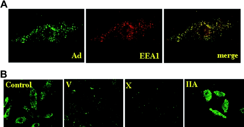
Figure 2. Immunostaining of adenoviral particles in BEAS-2B cells.
(A) Endocytosis of adenoviral particles into endosomes. BEAS-2B cells were incubated with adenovirus (MOI=20) for 6 h and were immunostained with anti-(adenoviral hexon) (left panel) and anti-EEA1 (middle panel) antibodies. A merged image is shown in the right panel. (B) Control BEAS-2B cells and cells expressing sPLA2-V, -X or -IIA were incubated with adenovirus (MOI=20) for 6 h and were immunostained with anti-adenoviral hexon antibody.

