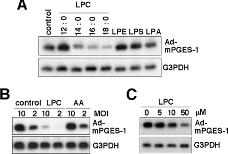
Figure 5. LPC blocks adenoviral infection into BEAS-2B cells.
(A) and (B) BEAS-2B cells were incubated with Ad-mPGES-1 at MOI=10 (A) and at MOI=2 or 10 (B) in the presence or absence (control) of various lysophospholipids or AA (each at 50 μM) for 24 h. In the case of LPC, molecular species having C12:0, C14:0, C16:0 and C18:0 at sn-1 were used. (C) The cells were incubated with Ad-mPGES-1 at MOI=10 in the presence or absence of the indicated concentrations of LPC with C18:0 at sn-1. The cells were then harvested and subjected to Northern-blotting with probes against mPGES-1 and G3PDH.

