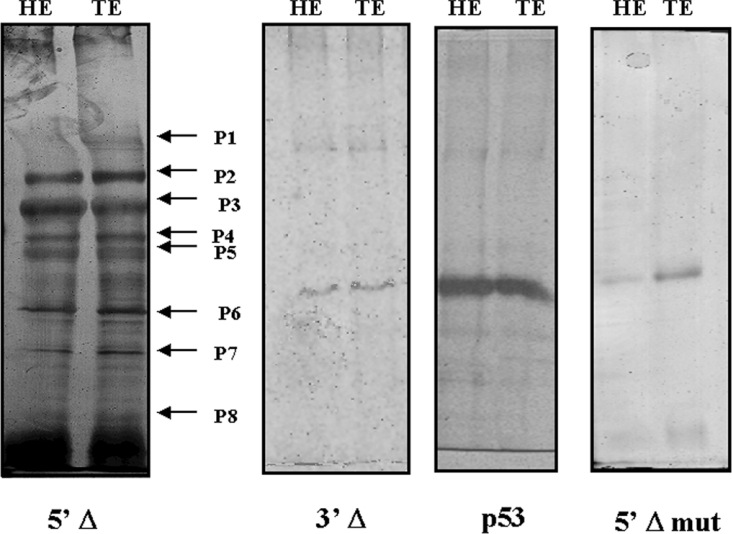
Figure 7. SDS/polyacrylamide gel of immunoprecipitation products.
Lanes named HE and TE correspond to reactions using 13.5 dpc embryo nuclear protein extracts from head and trunk respectively with anti-SMAD 1/SMAD 5/SMAD 8 antibody. The fragment used as a probe [5′-deleted fragment (5′Δ), 5′Δ mutated fragment (5′Δ mut) and 3′Δ fragment (3′Δ)] is indicated below each gel. The immunoprecipitated complex showed apparently the same composition using nuclear protein extracts from head or trunk. The arrows P1–P8 in the gel using 5′Δ fragment indicate the protein bands analysed by MS. The gel using 3′Δ fragment and 5′Δ mutated fragment did not show any immunoprecipitated protein. A control immunoprecipitation using an unspecific antibody (anti-P53) also did not show any protein.

