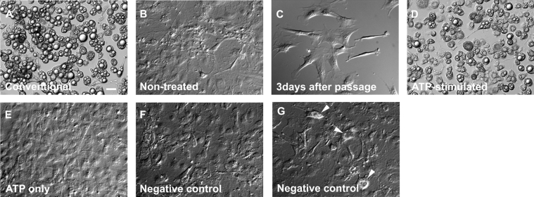
Figure 4. Effect of extracellular ATP on the differentiation of 3T3-L1 adipocytes.
DIC images of living cells. Differentiation protocols are described in the Experimental section. (A) Day 18, cells were treated with conventional protocol. (B) Day 18, cells were cultured for the same period as in (A) without adding adipogenic hormones and insulin. Cells in (A) and (B) were subcultured on the same day. (C) Day 3, 3 days after passage. (D) Day 18, cells were treated with the ATP-stimulated protocol. (E) Day 18, cells were solely exposed to 100 μM ATP on day 3 without adding adipogenic hormones and were then treated with insulin on day 6. (F and G) Day 18, cells were treated with the negative control protocol. Arrowheads in (G) indicate the cells containing lipid droplets. Cells in (C) to (G) were subcultured on the same day. Results are representative of six experiments. Scale bar, 20 μm.

