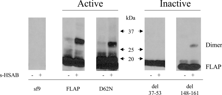
Figure 3. FLAP mutant dimers in Sf9 cells.
Sf9 cells were cultured and infected with baculoviruses. Post infection (3–4 days), Sf9 cells expressing the various FLAP mutants were sonicated and the lysates were treated with the cross-linking agent sulpho-HSAB (20 μg/ml) as described in the Materials and methods section. The reaction was stopped with the sample buffer and the proteins were analysed by SDS/PAGE using a 5–20% gradient and the FLAP antiserum anti-H5. Results shown are from one experiment and are representative of three different experiments.

