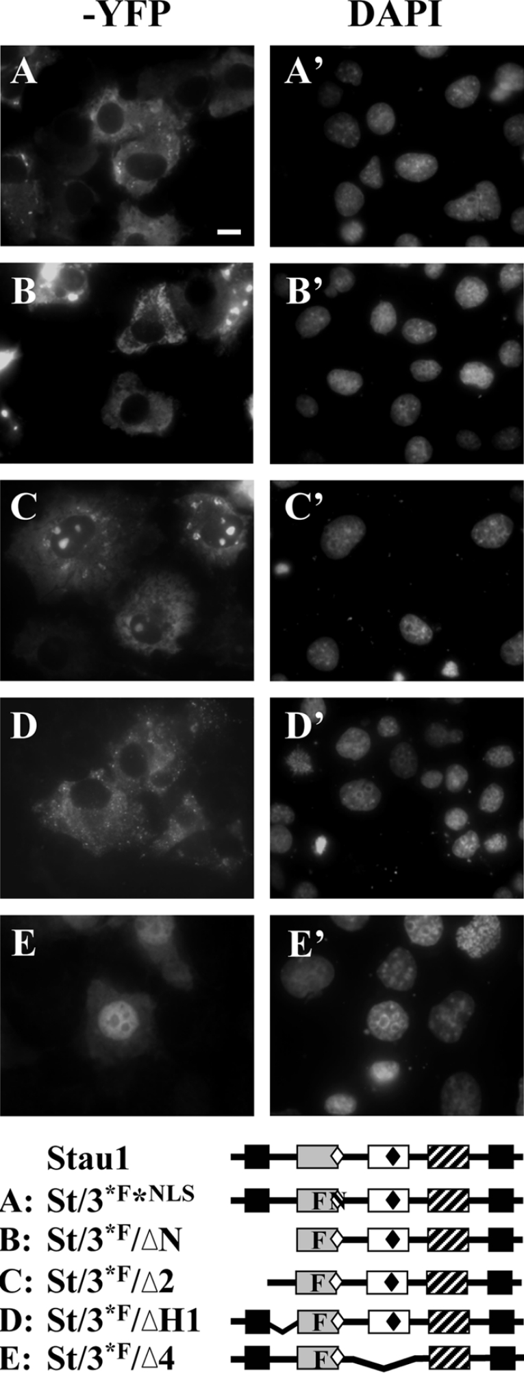
Figure 8. Involvement of the bipartite NLS and H1 regions in nuclear import of Stau1.
COS1 cells were transfected with 2 μg of cDNA coding for St/3*F*NLS–YFP (A, A′), St/3*F/ΔN–YFP (B, B′), St/3*F/Δ2–YFP (C, C′), St/3*F/ΔH1–YFP (D, D′) and St/3*F/Δ4–YFP (E, E′). Transfected cells were fixed 16 h post-transfection and the subcellular localization of each construct was monitored by YFP autofluorescence (A–E). Nuclei were stained with 300 nM DAPI solution (A′–E′). Scale bar, 20 μm.

