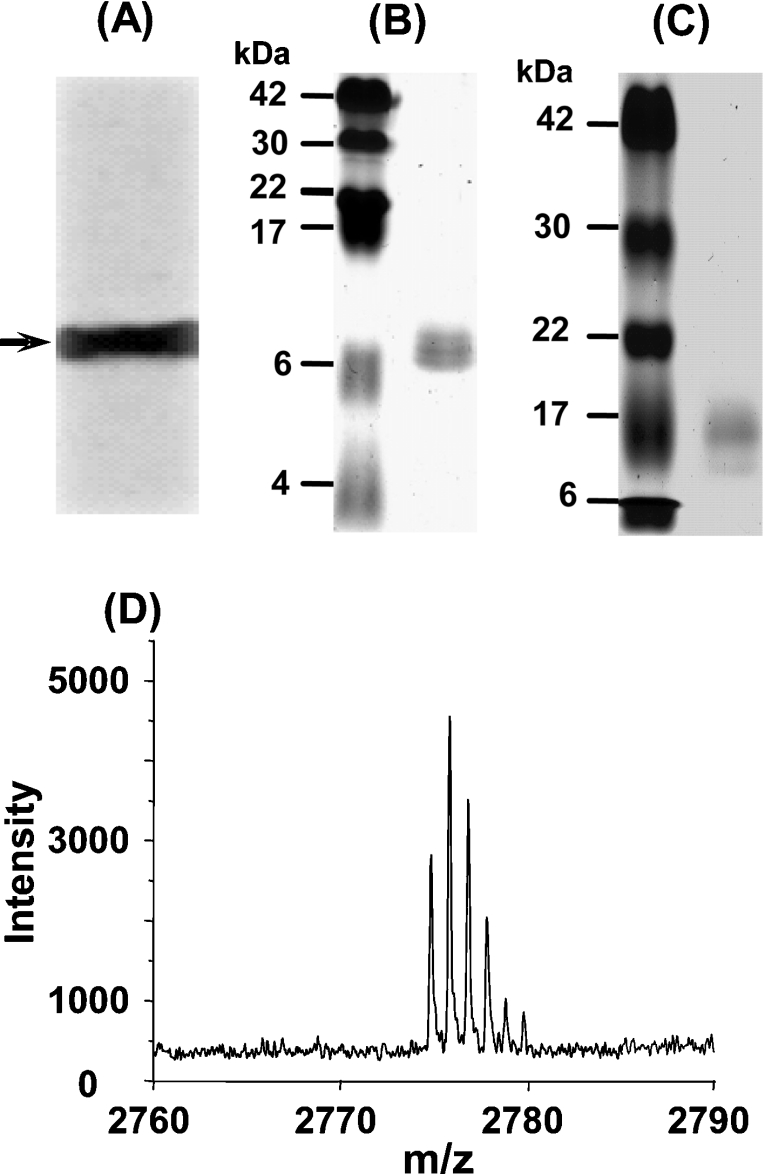
Figure 2. Analysis of expressed Hpn protein.
(A) Western blotting with His·Tag® antibody. The arrow indicates the position of the recombinant Hpn protein. (B) Purified Hpn subjected to SDS/20% PAGE. The left-hand lane contains the protein markers (sizes are given in kDa), and the right-hand lane contains purified Hpn protein, which migrates to approx. 7 kDa. (C) Purified Hpn subjected to SDS/15% PAGE. The left-hand lane contains the protein markers (sizes are given in kDa), and the right-hand lane contains purified Hpn protein, which migrates to approx. 16 kDa. (D) MALDI-MS spectrum of Hpn showing the major peak at m/z 2773.183 corresponding to the N-terminal fragment of recombinant Hpn.

