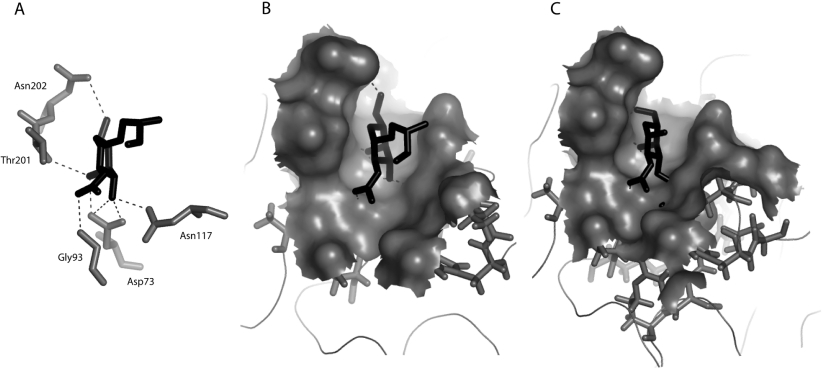
Figure 5. Docking of Tn and T antigen in the carbohydrate-binding site of Gleheda.
(A) Docking of Tn antigen in the carbohydrate-binding site. The network of hydrogen bonds (dark broken lines) connecting the sugars to residues Asp73, Gly93, Asn122, Trp117, Thr201 and Asn202 (Corey–Pauling–Koltun code) forming the monosaccharide-binding site of Gleheda. Residue His115 which stacks against the pyranose ring of the GalNAc is not shown. (B) Docking of GalNAcα1-Ser into the carbohydrate-binding cavity of Gleheda. (C) Docking of the T antigen into the carbohydrate-binding cavity of Gleheda showing the clash of the Gal residues with residues Asp73, His115 and Asn117. Both the carbohydrate-binding site and Tn or T antigen are similarly oriented in (A), (B) and (C). The Figure was drawn using PyMOL.

