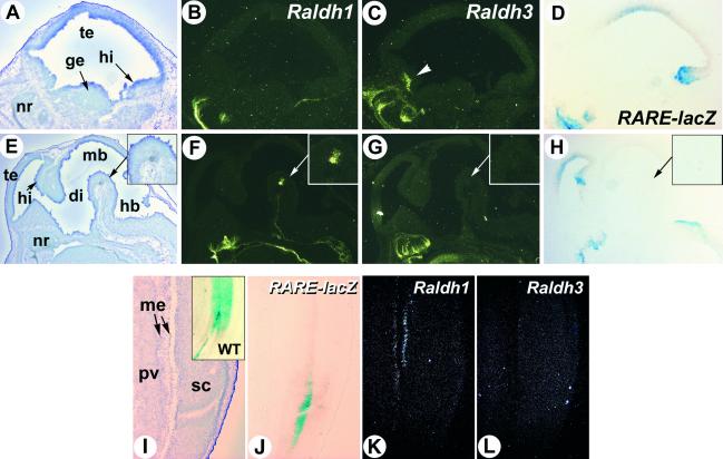
Fig 3.
RA reporter activity in the Raldh2−/− brain and spinal cord does not correlate with Raldh1 or Raldh3 gene expression. (A–H) Sagittal sections through the lateral region of the forebrain (A–D) and the near-midline brain structures (E–H) of an E14.5 transgenic Raldh2−/− embryo were hybridized with Raldh1 (B and F) and Raldh3 (C and G) riboprobes, or X-Gal-stained (D and H). A higher magnification of the ventral midbrain Raldh1-expressing area is shown (Insets) (arrows in E–H). Raldh3-specific expression in the rostro-ventral telencephalon is indicated by an arrowhead. (I–L) Serial sections through the lumbar spinal spinal cord of an E14.5 transgenic Raldh2−/− embryo were X-Gal-stained (J) or hybridized with Raldh1 (K) and Raldh3 (L) riboprobes. (I Inset) The X-Gal pattern obtained in a WT spinal cord. di, diencephalon; ge, ganglionic eminence; hb, hindbrain; hi, hippocampal region; mb, midbrain; me, meninges; nr, nasal region; pv, prevertebrae; sc, spinal cord; te, telencephalon. (Magnifications: ×5, E–H; ×8, A–D; ×10, I–L.)

