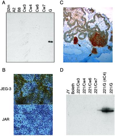
Fig 1.
Characterization of HLA-G-specific mAbs. (A) Western blot of bacterially produced solubilized inclusion bodies from various soluble recombinant HLA molecules detected by MEM-G/1 mAb. (B) Cytospin preparations of the HLA-G+ choriocarcinoma cell line JEG-3 and the HLA-G− cell line JAR were stained with the MEM-G/1 mAb. Whereas JAR is completely negative, HLA-G staining (brown) can be seen clearly in the JEG-3 sample. Note that not all of the JEG-3 cells stain positively, which is consistent with the heterogeneous nature of this tumor line. (C) Immunohistochemistry of paraffin-embedded placental sections using the MEM-G/1 mAb. HLA-G staining (dark brown) was detected on the cell islands (denoted by arrows) that had migrated from the villi. (D) The MEM-G/11 mAb was used to immunoprecipitate HLA-G from surface-biotinylated B-lymphoblastoid cell lines and from various class I MHC transfected into the MHC-deficient 721.221 B-lymphoblastoid cell line. ECL visualization demonstrated that only the two different clones of HLA-G+ transfectants were recognized.

