Fig 1.
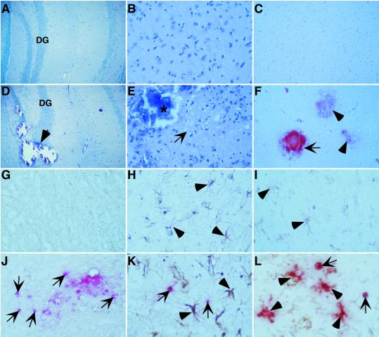
Severe pathological changes in the brain of GIFN12 STAT1-null mice. Brain from 3-month-old GIFN12 (A–C) or GIFN12 STAT1-null (D–F) mice is shown. Luxol fast blue stains (A and D) showed mineral deposits (D, arrow) in the dentate gyrus (DG) region of the hippocampus, causing marked disruption of the dentate granule neurons in the GIFN12 STAT1-null specimen (both show original magnification, ×100). Hematoxylin/eosin stains (B and E) showed gross loss of neurons and spongiform changes (E, arrow) in proximity to the mineral deposits (*) in the basal ganglia region from this GIFN12 STAT1-null specimen (B and E, original magnification, ×400). Alizarin Red S-stained (C and F) advanced (F, arrow) as well as early (F, arrowheads) mineral deposits in brain from GIFN12 STAT1-null mice (C and F, original magnification, ×200) are shown. TUNEL staining revealed markedly increased cell death (arrows) in the brain from GIFN12 STAT1-null mice (J–L) compared with GIFN12 (G–I) animals. TUNEL staining combined with cell marker-specific staining for neurons (neurofilament; G and J), astrocytes (GFAP; H and K, arrowheads), and macrophage/microglia (tomato lectin; I and L, arrowheads) is shown. The region shown in G–L is the basal ganglia at an original magnification of ×600.

