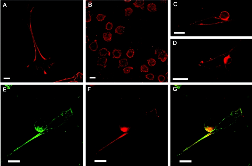
Figure 5. Subcellular location of GM1 and GM-CSF-Rα in cell cultures and co-culture systems.
GM1 was examined by incubation of cholera toxin, followed by a monoclonal anti-(cholera toxin) antibody or by the fluorescence of FITC-conjugated cholera toxin. Cells were immunostained for GM-CSF-Rα with a polyclonal anti-(GM-CSF-Rα) antibody. (A) AFT-024 cells immunostained for GM1. (B) FDC-P1 cells immunostained for GM1. (C–E) Co-cultures immunostained for GM1. (F) Co-cultures immunostained for GM-CSF-Rα. (G) Merged images from (E) and (F). Single confocal sections of 0.7 μm were taken parallel to the coverslip. Scale bars, 10 μm.

