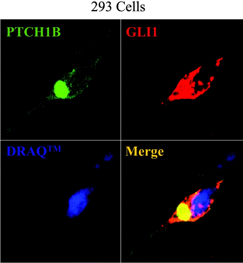
Figure 3. Subcellular localization of co-transfected PTCH1 and GLI1.
HEK-293 cells were transfected with FLAG-tagged PTCH1 with HA-tagged GLI1 and stained with antibodies to the respective tag. The proteins were visualized by immunofluorescence confocal microscopy. Dual-channel imaging indicates co-localized region, with GLI1 shown in red and PTCH1 in green. Nuclear staining with DRAQ™ is shown in blue.

