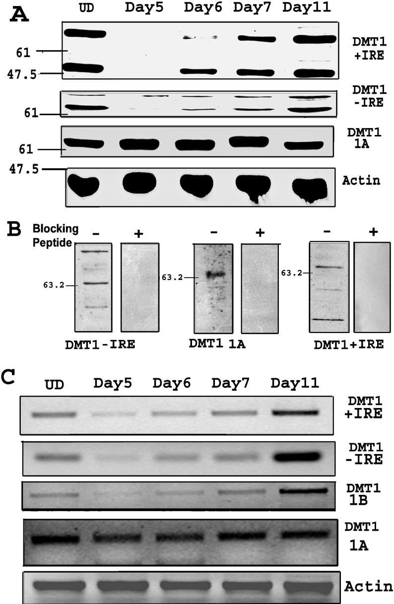
Figure 1. Down-regulation of specific isoforms of DMT1 during neural differentiation.
(A) Western-blot shows down-regulation of +IRE and −IRE isoforms of DMT1 on day 5, a day after removal of RA. The expression of +IRE and −IRE DMT1 isoforms is recovered and is similar to the UD (undifferentiated cells) level by day 11. Western-blot using anti-1A antibody shows no significant change during differentiation. Anti-β-actin antibody was used as gel loading control. Protein loading was 15 μg/lane and electrophoresis was on 12% polyacrylamide gel. (B) Western-blot showing preincubation of antibody with the respective blocking peptide results in inhibition of all the bands on the blot, indicating specificity of the bands. (C) RT-PCR of cells during differentiation shows decrease in +IRE, −IRE and 1B DMT1 mRNA on day 5 after removal of RA. The mRNA expression recovered by the seventh day (day 11) after differentiation was initiated by removal of RA. RT–PCR for the 1A isoform shows no change during differentiation. RT-PCR for β-actin was done as the RNA control. Total RNA (500 ng) was used for each RT-PCR assay. PCR was performed for 30 cycles in an Eppendorf Mastercycler and the products were electrophoresed on a 2% (w/v) agarose gel.

