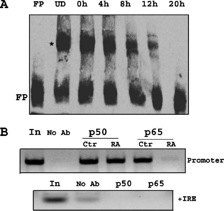
Figure 9. NF-κB binding to the promoter region of the 1B isoform of DMT1 is decreased after removal of RA.
(A) EMSA was performed using nuclear extracts from cells (undifferentiated and 0, 4, 8, 12 and 20 h after removal of RA). Biotin-labelled probe representing promoter region of 1B isoform was incubated with nuclear extracts, and using LightShift chemiluminescent EMSA kit (Pierce), gel-shift was observed. There was constitutive binding of nuclear factor (denoted by *) in nuclear extracts from undifferentiated cells (UD). There was a decrease in binding (less gel-shift band intensity) after removal of RA, also seen by an increase in free probe at the bottom (FP). (B) ChIP assay using primers specific for 1B promoter region shows constitutive binding of p50 and p65 in undifferentiated cells. There was no change in the binding of p50, 10 h after RA removal (RA), but there was a decrease in binding of p65. The first lane (In, input) indicates total chromatin DNA used in PCR reaction, while as a control, no antibody control was used in immunoprecipitation reaction (No Ab). As a PCR control, same samples were used for PCR using +IRE primers. Bands are seen only in the first lane and the no antibody lane.

