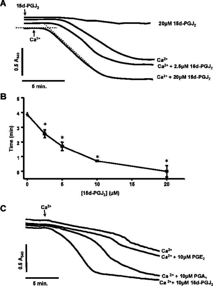
Figure 1. Ca2+-induced pore opening is accelerated by 15d-PGJ2 in a concentration-dependent manner.
Mitochondria (1 mg/ml) were incubated in the spectrophotometer in swelling buffer with succinate (5 mM) and rotenone (1 μM). (A) Where indicated, increasing concentrations of 15d-PGJ2 were present from the beginning of the experiment and Ca2+ (10 μM) was added after 2 min. (B) Lag time (after the addition of Ca2+) was measured by determining the intercepts of minimum and maximum rates of change in absorbance as shown in (A) for the Ca2++15d-PGJ2 sample (20 μM). Results are expressed as the means±S.E.M. for at least three experiments. *P<0.001 versus 0 μM 15d-PGJ2. (C) Prostaglandins A (10 μM), E (10 μM) and J2 (10 μM) were present from the beginning of the experiment and Ca2+ (5 μM) was added at 2 min. Data are single traces representative of 5–7 individual experiments.

