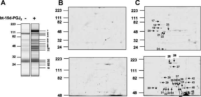
Figure 11. Identification of the mitochondrial electrophile-responsive proteome.
Isolated mitochondria were incubated in the presence or absence of 20 μM bt-15d-PGJ2 for 15 min at 25 °C, and labelled proteins were affinity precipitated with neutravidin, as described in the Experimental section, and visualized using 1D SDS/PAGE followed by Coomassie Blue staining (A). Affinity-precipitated proteins from untreated mitochondria (B) and from bt-15d-PGJ2-treated mitochondria (C) were also separated using 2D SDS/PAGE and stained for protein. Higher-molecular-mass proteins were optimally resolved using longer run times (B and C, lower panels).

