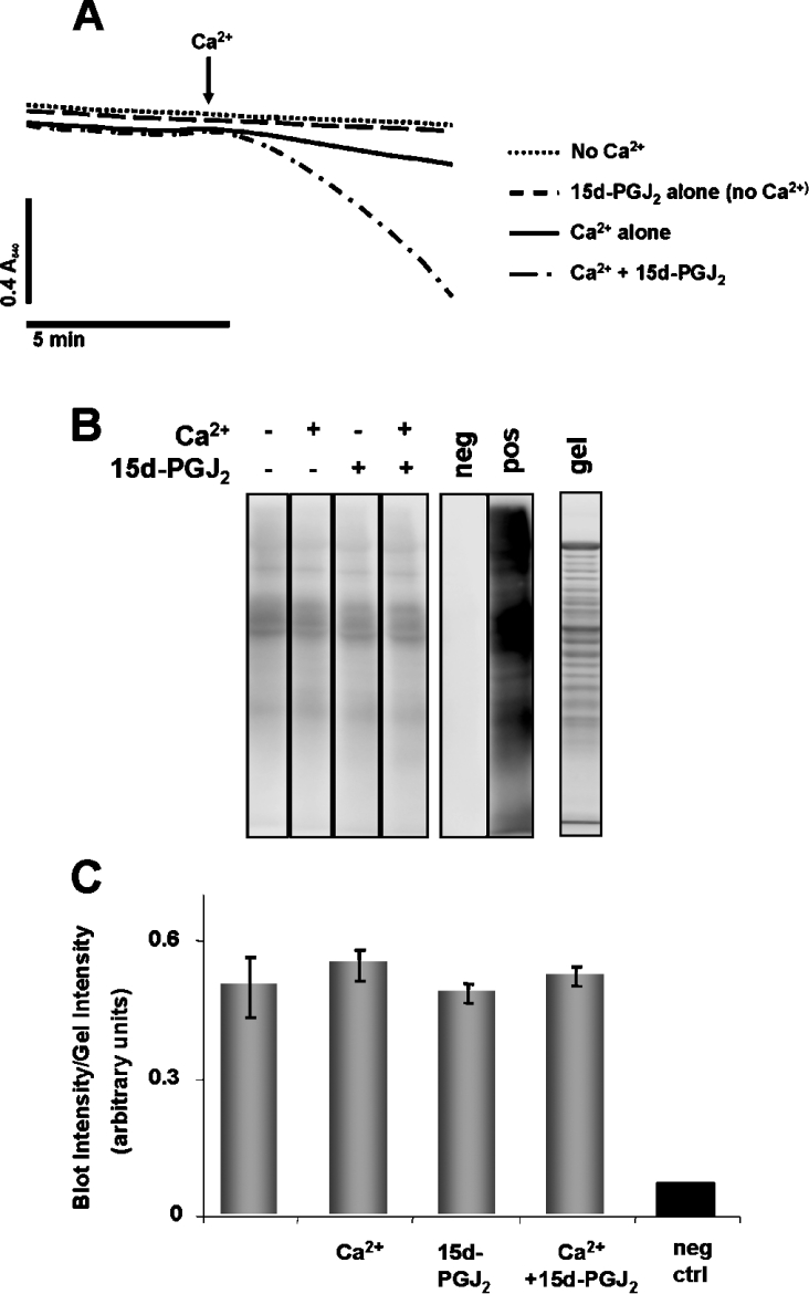
Figure 6. Detection of protein carbonyl formation on mitochondrial proteins after exposure to Ca2+ and 15d-PGJ2.
(A) Mitochondria (1 mg/ml) were incubated in swelling buffer with succinate (5 mM) and rotenone (1 μM) with or without 80 μM Ca2+ or 20 μM 15d-PGJ2 for the time period shown before samples were flash frozen in liquid nitrogen. (B) Carbonyl formation on mitochondrial proteins was detected after derivatization with DNPH followed by Western blot analysis, as indicated. The negative control (neg) was a sample of mitochondria in which the DNPH derivatization step was omitted. The positive control (pos) was prepared by incubating mitochondrial proteins with iron/ascorbate. Proteins separated on an identical gel were stained with Sypro Ruby (gel). (C) Quantitation of the Western blot in (B). Total density from each lane on the Western blot was normalized to the total density of the corresponding lane on the gel. Results are expressed as the means±S.E.M. (n=3).

