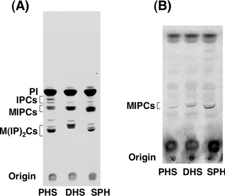
Figure 3. Synthesis of complex sphingolipids in LCB-reconstituted cells.
(A) [3H]myo-inositol labelling of LCB-reconstituted cells. TMY85 cells cultured with 5 μM PHS, DHS or SPH were labelled with [3H]myo-inositol for 5 h at 30 °C. Incorporated radioactivity was quantified, and equivalent samples (c.p.m.) were used for further analysis. Lipids were extracted and separated by TLC with chloroform/methanol/4.2 M ammonia (9:7:2, by vol.) as the solvent system. (B) Detection of unlabelled MIPCs in LCB-reconstituted cells. TMY85 cells (3.0 D600 units/ml) were cultured with PHS, DHS or SPH as in (A) and collected. Lipids were extracted, separated by TLC and then visualized by orcinol/H2SO4 reagent.

