
Figure 7. Cytochemical demonstration of iron by the sulphide-silver method (autometallography).
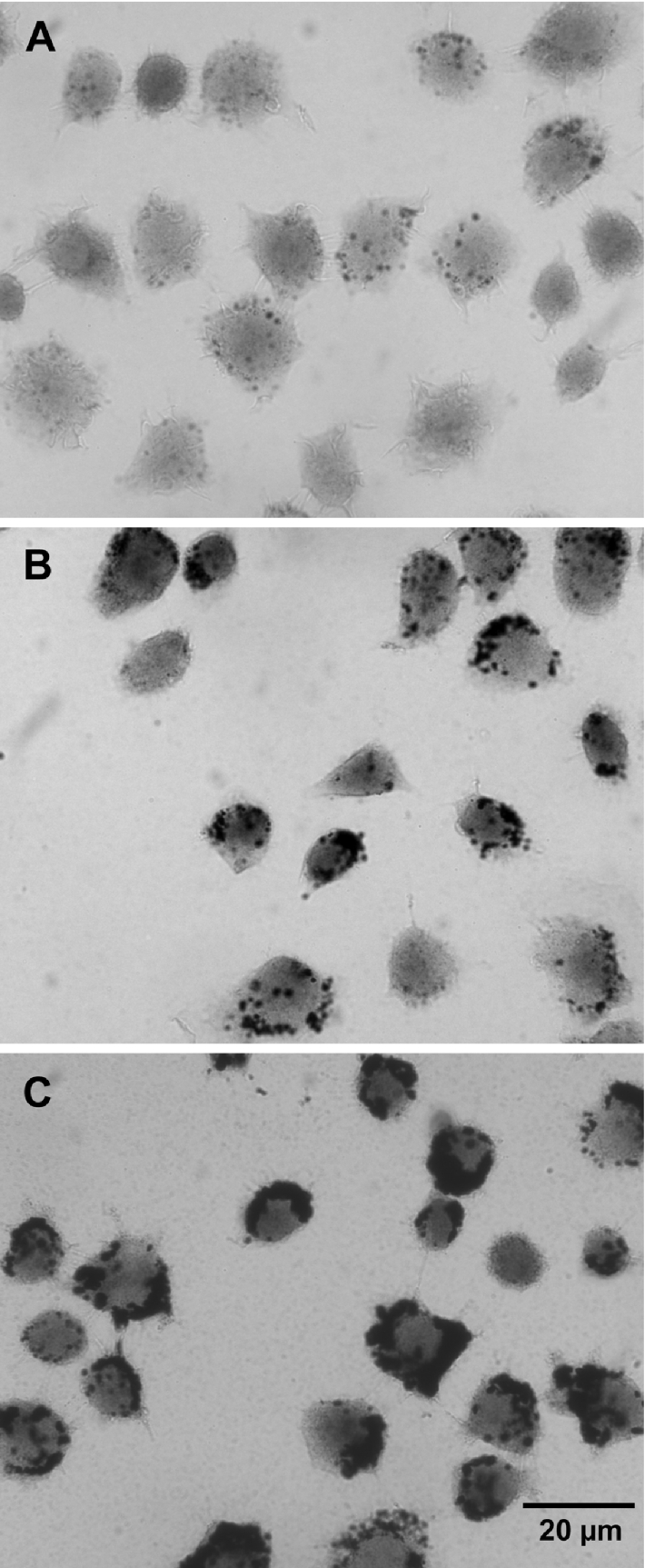
Cells showed a distinct lysosomal pattern of black, granular silver precipitates, indicating the presence of concentrated lysosomal low-molecular-mass iron. In control cells (A), only a few granules were found, reflecting a limited number of autophagolysosomes with Fe-containing macromolecules under degradation, while cells exposed for 3 h to a hydrated iron phosphate complex [obtained by adding FeCl3 to the culture medium to final concentrations of 30 μM (B) or 100 μM (C)] showed considerably more granules, reflecting fluid-phase endocytosis into late endosomes/lysosomes of the Fe complex. There was also a weak (A) or more substantial (B, C) yellowish diffuse colorization, reflecting the occurrence of cytosolic low mass iron (development time=50 min).