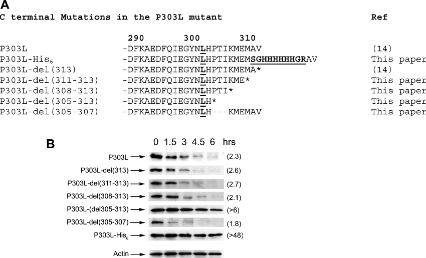
Figure 6. Degradation of the P303L enzyme is determined by its C-terminal region.
(A) Amino acid sequences of the various C-terminal mutants of the P303L form of TS are shown. The P303L substitution and His6 insertion are indicated in boldface and underlined. Additional residues flanking the His6 sequence (Ser-Arg and Gly-Arg) occur as an inevitable consequence of insertion of a NotI restriction site for subcloning (see the Experimental section), and are also indicated in boldface and underlined. (B) RJK88.13 cells were transiently transfected with plasmids encoding the P303L mutant and its variants P303L-del(313), P303L-del(311–313), P303L-del(308–313), P303L-del(305–313), P303L-del(305–307) and P303L–His6. After 24 h, transfected cells were split into 60 mm plates, and cycloheximide treatment (50 μg/ml) was initiated 24 h later. Cells were harvested after the indicated times of cycloheximide exposure, and steady-state levels of the P303L polypeptide were determined by Western-blot analysis of total cell extracts, using the human TS monoclonal antibody. As a control, immunoblots were stripped and steady-state levels of actin were analysed using an α-actin monoclonal antibody. A representative blot is shown (Actin) to indicate equal loading. Half-lives (h), estimated by scanning and quantification of the films, are indicated in parentheses.

