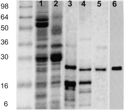
Fig 1.
Purification of PsbS. Silver-stained SDS polyacrylamide gel showing spinach thylakoids (lane 1), PSII membrane fragments (lane 2), pellet after extraction with DM (lane 3), supernatant after extraction with cholate (lane 4), and after passing down a Sephadex G-25 column (lane 5). A Western blot of lane 5 with anti-PsbS antibody is shown in lane 6. Molecular weight markers (See Blue, Invitrogen) are shown in the far-left lane.

