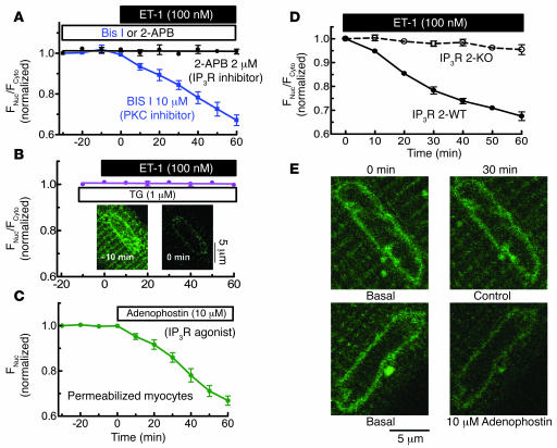
Figure 3.
ET-1–induced HDAC5 nuclear export is dependent on InsP3R and Ca2+ in InsP3-sensitive stores. (A) HDAC5-GFP–expressing myocytes were pretreated with 2 μM 2-APB (n = 6) or 10 μM Bis I (n = 5) for 30 minutes, followed by application of 100 nM ET-1. (B) SR and nuclear envelope Ca2+ stores were depleted by preincubation with SR/ER Ca2+-ATPase (SERCA) inhibitor TG 1 μM (n = 7) for 10 minutes. Insets are the Fluo-5N images, showing that this TG treatment depleted nuclear envelope Ca2+ stores. (C) In permeabilized HDAC5-GFP expressing myocytes, 10 μM adenophostin was applied after 30 minutes at 100 nM [Ca2+]i (n = 8). HDAC5 nuclear export was measured as the decrease of nuclear fluorescence. In the permeabilized cell, cytosolic concentration is irrelevant as HDAC5-GFP can readily diffuse to the bath. (D) ET-1–induced HDAC5 nuclear export assessed in mouse ventricular myocytes that lack InsP3R2 (InsP3R 2 KO) or WT littermates. (E) Fluo-5N–loaded myocytes were permeabilized and treated without or with 10 μM adenophostin for 30 minutes. Nuclear envelope Ca2+ release was indicated by decreased Fluo-5N fluorescence.

