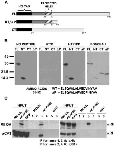
Figure 4.
Analysis of PKA binding to AKAP28. (A) Schematic representation of full-length and truncation His6-AKAP28 proteins. All proteins are drawn to scale and the amino acid numbers are shown. (B) Equal moles of His6 fusion proteins were resolved by SDS-PAGE, transferred to PVDF membrane, probed with biotinylated RII, subsequently incubated with streptavidin-HRP, and developed with ECL. Biotinylated RII was either preblocked with no peptide, 1 μM competitive Ht31 or 1 μM negative control Ht31PP. A representative Ponceau-stained membrane is also shown. (C) HeLa cells were either mock infected (MOCK) or infected with a control adenovirus expressing GFP, or an adenovirus expressing both GFP and HA-tagged AKAP28 (AKAP28). Cells lysates (300 μg) were incubated with monoclonal HA.11 or mouse IgG1a. Immunocomplexes were collected on protein G and subsequently analyzed by Western blot or RII overlay as indicated. Inputs (one-fifth of total) for all cell lysates are shown. Lanes 1, 3, and 5 were incubated with HA antisera, whereas 2, 4, and 6 were incubated with mouse IgG1a. All data are representative of multiple independent experiments.

