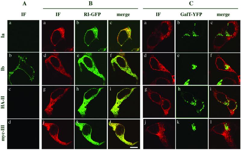
Figure 2.
Only UPIb is expressed on the cell surface. Expression of the individual uroplakin subunits in 293T cells was carried out as described in MATERIAL AND METHODS. An HA-tagged form of UPII (HA-UPII) or a myc-tagged form of UPIII (myc-UPIII) was used because antibodies against UPII or UPIII were not available for immunostaining. (A) Transfected cells were immunostained under nonpermeabilized condition. fluorescein isothiocyanate-conjugated donkey anti-rabbit IgG (a and b) or Texas Red-conjugated donkey anti-mouse IgG (c and d) was used. Only UPIb was stained at the plasma membrane. (B) To determine the intracellular location of individual uroplakins, 293T cells were cotransfected with the ER marker ribophorin I-GFP (a–l) and UPIa (a–c), or UPIb (d–f), or HA-UPII (g–i) or myc-UPIII (j–l). Cells were permeabilized by adding 0.05% saponin to the blocking solution and immunostained. All the secondary antibodies were conjugated with Texas Red. Uroplakins retained intracellularly colocalized with RI-GFP. (C) Cells were cotransfected with the Golgi marker galactosyl transferase tagged with YFP (GalT-YFP; a–l) with UPIa (a–c), or UPIb (d–f), or HA-UPII (g–i) or myc-UPIII (j–l). No significant colocalization between GalT-YFP and uroplakins was observed. All the immunostained cells were analyzed by confocal microscopy.

