Figure 2.
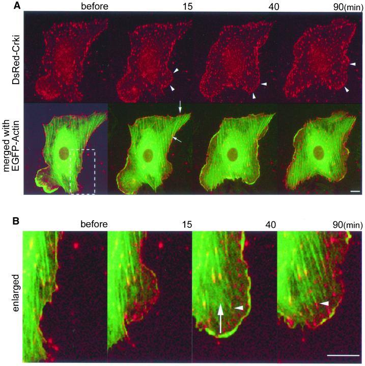
CrkI is translocated to nascent focal complexes upon ephrin-B1 stimulation and is involved in the development of focal adhesion. HAECs cultured on a collagen-coated glass-base dish were transfected with pCA-DsRed-CrkI, starved for 8 h, and stimulated with preclustered 1 μg/ml soluble ephrin-B1/Fc. EGFP-actin and DsRed-CrkI were imaged on an IX-70 inverted microscope (Olympus). A series of DsRed and EGFP images were collected by MetaMorph 4.6 software. Before, before the stimulation; 15, 40, and 90 min, time for stimulation. Bar, 10 μm. (A) Arrowheads indicate typical accumulation of DsRed-CrkI to nascent focal complexes (top). The GFP image of the same cell transfected with pEGFP-actin was merged with the DsRed-CrkI image (bottom). Arrows indicate an example of focal adhesions connecting actin stress fiber (also see Video 2A2). (B) The boxed region in the lower panel of A was enlarged. An arrow indicates a focal complex detached from the substratum and floating in membrane ruffles. The arrowhead indicates the development of a focal complex to a focal adhesion (also see Video 2B).

