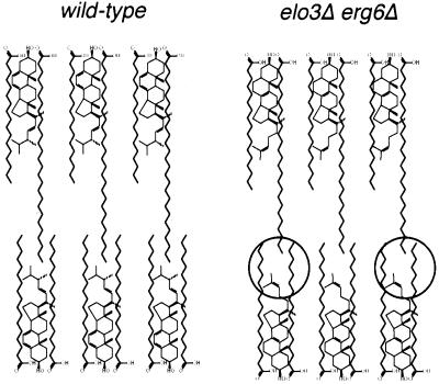
Figure 12.
Comparison of the structure of the plasma membrane of wild-type and elo3Δ erg6Δ mutant cells. Schematic model of the presumed structure of the hydrophobic core of the plasma membrane of wild-type cells compared with cells that contain C22-substituted sphingolipids and zymosterol instead of ergosterol. Alteration in the hydrophobic core of the mutant membrane is encircled. See text for details.

