Figure 1.
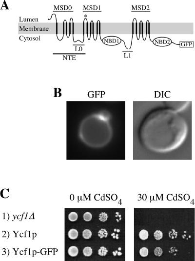
GFP-tagged Ycf1p localizes to the vacuolar membrane and is functional. (A) Proposed topology of yeast Ycf1p is shown. The core region contains two membrane spanning domains (MSD1 and MSD2) and two nucleotide binding domains (NBD1 and NBD2), separated by a linker (L1) (note, for simplicity sake L1 is omitted in subsequent figures). The NTE is comprised of MSD0 and L0. The proposed topology for Ycf1p is modeled after the experimentally determined topology of the Ycf1p human homologue MRP1 (Bakos et al., 1996; Hipfner et al., 1997). Any small differences between Ycf1p and MRP1 would be unlikely to influence the conclusions of this study. The GFP coding sequence is fused immediately before the stop codon of Ycf1p. (B) GFP fluorescence pattern (left) and corresponding DIC image (right) of cells expressing Ycf1p-GFP are shown. The vacuole appears as an indentation in the DIC images. The brighter area of fluorescence appears to correspond to a region where two vacuolar lobes intersect. (C) Cadmium resistance was tested by spotting an aliquot (5 μl) of cells at 0.1 OD600 and serial 10-fold dilutions thereof onto plates containing 0 or 30 μM CdSO4 and incubating at 30°C for 3 or 6 d, respectively. The strains used are SM4516 (row 1), SM4517 (row 2), and SM4518 (B and row 3).

