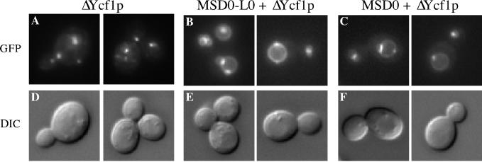
Figure 7.
L0 is not necessary for the vacuolar localization of Ycf1p. The GFP fluorescence pattern of the C-terminal partial molecule ΔYcf1p is shown for cells expressing solely ΔYcf1p (A), for cells coexpressing ΔYcf1p and MSD0-L0 (B), or for cells coexpressing ΔYcf1p and MSD0 (C). Two examples are shown for each. The corresponding DIC images of these cells are also shown (D–F). The strains used are SM4764 (A), SM4742 (B), and SM4778 (C).

