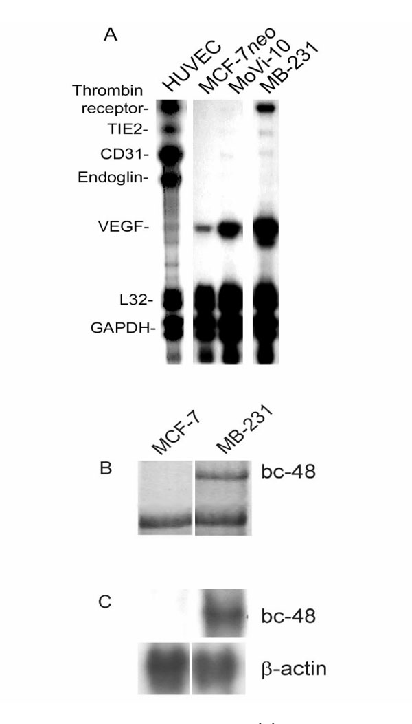
Figure 1.

(A) Ribonuclease protection assay analysis, (B) differential display analysis, and (C) Northern blot analysis of vascular markers expressed by human breast cancer cell lines, including the following: thrombin receptor, endothelin B receptor (methods described by Kirschmann et al [43]), TIE2, CD31, endoglin and VEGF. Equal loading was assessed by ribosomal protein L32 (L32) and glyceraldehyde-3-phosphate dehydrogenase (GAPDH) expression (A), genes not differentially expressed (B), and β-actin (C). These data are summarized in Table 1.
