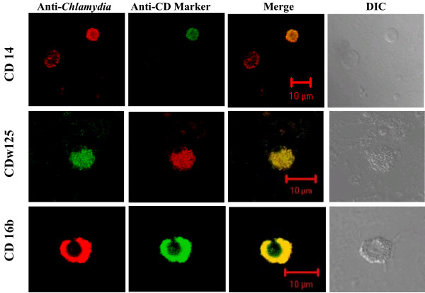
Figure 3.
Infected and uninfected WBCs dual immunostained with CD Specific monoclonal and anti-Chlamydia antibody. Representative medial confocal microscopic images from the Z-axis series of optical sections through Chlamydia-infected human PBMC cells. Immunostaining used anti-Chlamydia EB antibody and either anti-CD14(monocyte), anti-CD 16b(neutrophil), or anti-CDw125 (eosinophil/basophil) mAb as indicated. Respectively, the Merge and DIC columns show the combined FITC and TRITC images, and the distribution and morphology of the cells in the field. Note: In vivo each of the three cell types can be infected by Chlamydia and the CD14 row includes a Chlamydia infected cell (TRITC positive) that is not immunostained by the ant-CD14 monoclonal (FITC negative).

