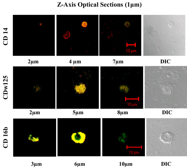
Figure 4.
Merged images at different depths from the confocal Z-axis series of dual immunostained WBCs. These merged TRITC-FITC confocal images are from different depths in the same series of Z-axis optical sections acquired for each of the cell types shown in Figure 3. The position of individual sections relative to the cell surface is indicated in μm below each image. Optical section images were acquired with the Zeiss Metaconfocal System.

