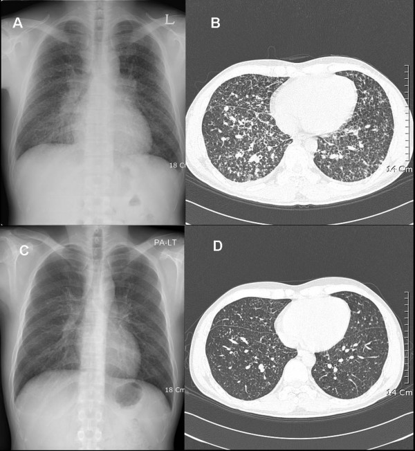
Figure 1.
Chest X-ray (CXR) and high-resolution computed tomography (CT) findings on admission, and again at five months following combined treatment. Top (on admission): (A) Posterior-anterior chest radiograph (P-A CXR) shows multiple tiny reticular nodules on both lungs; (B) Tiny centrilobular and subpleural nodules are evident in both lungs on the chest CT. Bottom (five months after combined treatment): (C) A follow-up CXR shows improvement in the multiple reticular nodules; (D) A follow-up CT scan reveals the improvement of the residual nodules.

