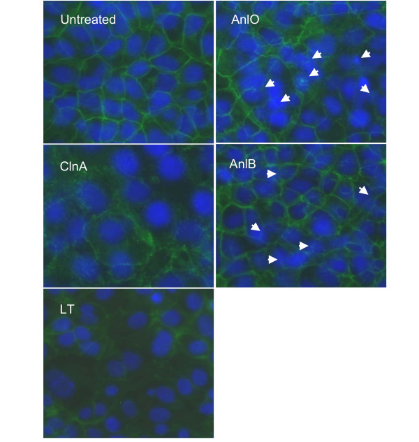
Figure 4.

Immunohistochemical detection of E-cadherin in monolayers of NMuMG cells after treatment with LT and hemolytic proteins (1 μg/ml each for 16 h). Nuclei are stained blue with DAPI, and E-cadherin is stained green with FITC-conjugated anti-E-cadherin antibody. In the case of AnlO and AnlB, arrows indicate some of the most damaged areas. 40× magnification.
