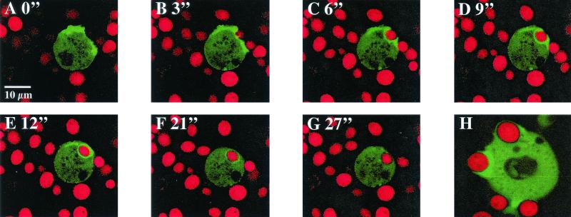
FIG. 4.
Distribution of a GFP-actin fusion protein during phagocytosis. AX2 wild-type cells and 1a1 mutant cells expressing a GFP-actin fusion protein were fed with TRITC-labeled yeast cells. Phagocytosis was observed by confocal laser scanning microscopy at the indicated times (in minutes). GFP-actin distribution did not differ between the two Dictyostelium strains. Thus, we only show the series of confocal images for the mutant 1a1. (A and B) Enrichment of actin beneath the cell surface at sites of contact with the yeast particle. (C and D) GFP-actin-stained leading edges begin to engulf the yeast cell. (E) The engulfed yeast cell is surrounded by a ring of GFP-actin. (F and G) GFP-actin disassembled from the mature phagosome. (H) AX2 cell forming a phagocytic cup which shows an enrichment of F-actin.

