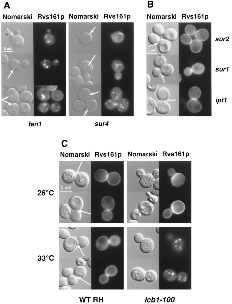
FIG. 7.
Rvs161p-GFP localization is impaired in fen1, sur4, and lcb1-100 mutants. Different mutant cells grown in SD-casa medium were observed by Nomarski and fluorescence microscopy to visualize whole cells and Rvs161p-GFP localization, respectively. (A) In the fen1 and sur4 mutants, the arrow on the Nomarski images indicates the structures that contain Rvs161p-GFP. (B) In the sur2, sur1, and ipt1 mutants, the arrow on the Nomarski images indicates the septum between the mother cell and the bud. (C) Rvs161p-GFP localization in the lcb1-100 mutant and the corresponding wild-type (RH1800) cells grown at 26°C and shifted to 33°C for 90 min. The arrow on the Nomarski images indicates the septum between the mother cell and the bud.

