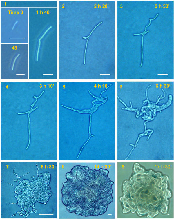
Figure 9.
Growth of SINett on agar as seen at the light microscope. In this SIN mutant a bent appears at an early stage of colony formation (1) and a double rupture occurs (2). From these ruptures 3 parallel filaments start growing (3). In (4) new ruptures in the upper filament appear. In (5–6) many filaments aggregate in bundles covering the central part of the starting filament. Note that magnification is the same in these 6 figures. In (7) the colony covers almost all of the central space. In (8–9) the final shape is shown. Several layers of cells are superimposed. They appear more evident in (9) where the picture was taken with a filter. Scale bar: 1–6 = 20 μm, 7–9 = 100 μm.

