Figure 7.
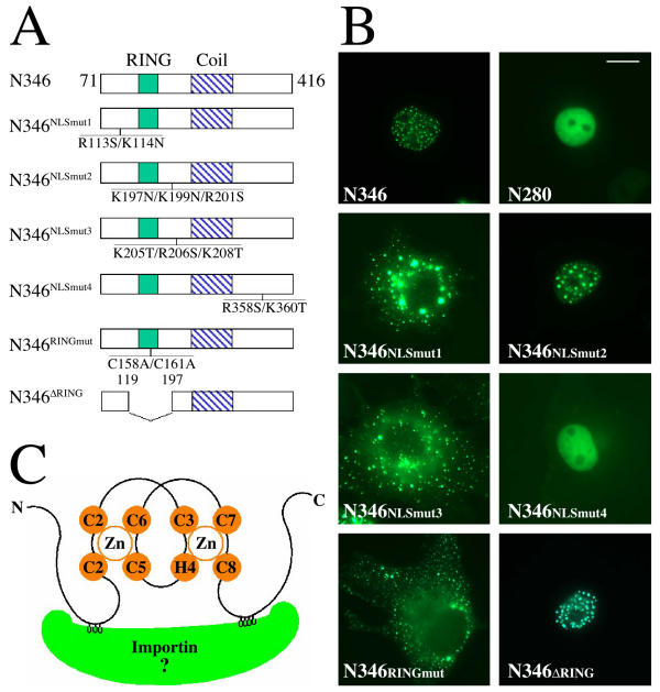
Mapping the nuclear localization signal of mammalian COP1 protein (A) Diagrams of the point-mutation and deletion-mutation constructs in GFP-N346 in comparison to the wild-type GFP-N346. The position and the amino acid replacements in each mutant protein are indicated. (B) GFP fluorescence study of the subcellular localizations of the mutant GFP-N346 proteins. The studies were carried out in COS7 cells. The same magnification was used for all images and the scale bar for 10 micrometer was indicated in the N280 image. (C) Schematic illustration of COP1 RING finger bridged bipartite nuclear localization signal (NLS) binding to importin. The two zinc atoms and zinc binding residues of COP1 RING finger are shown in orange. The two clusters of positively charged residues of COP1 NLS (in grey) bind to two binding pockets on the inner surface of importin (in green).

