
Figure 2. Identification of the sst1candidate gene.
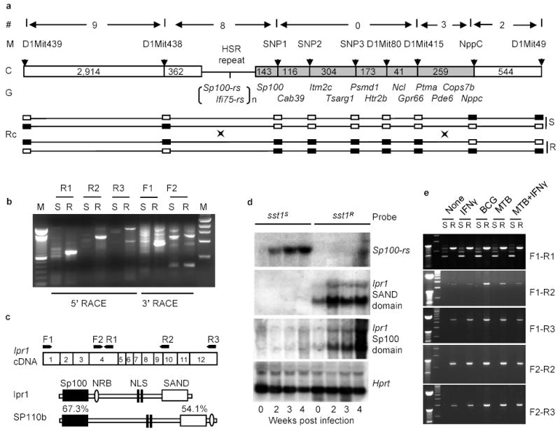
a, Physical map of the sst1 minimal region. (#)- number of recombination evens, (M) – polymorphic markers, (C) – chromosome with distances between the markers (kb); (G) – known genes; (RC) - recombinant chromosomes containing the sst1 resistant (R) or susceptible (S) alleles, genotypes for each marker are represented by solid (B6) and opened (C3H) boxes; b, Analysis of the Ifi75-rs expression in the tuberculosis lung lesions of the sst1 congenic mice by RACE. c, Domain structure of the Ipr1 and its human homolog SP110b and location of the PCR primers. d, Ipr1 and Sp100-rs gene expression in the lungs during MTB infection (Northern blot); e, Ipr1 gene expression in sst1S (S) or sst1R (R) macrophages infected MTB, BCG or activated with IFN-γ in vitro.