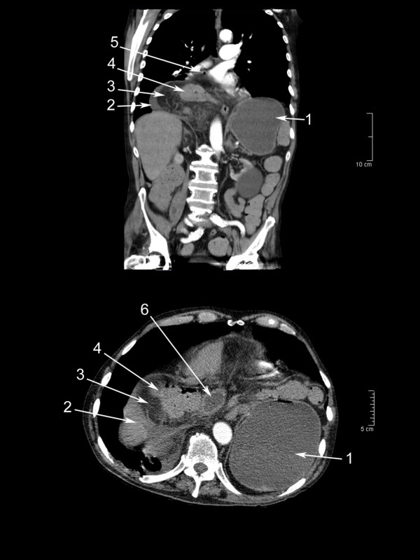
Figure 1.

Computed tomography on the day of arrival. Frontal and transverse projections showing: 1. Intraabdominally distended gastric fundus. 2. Fluid in hernia. 3. Herniated major omentum. 4. Herniated transverse colon. 5. Small bubble of free air, not noticed primarily. 6. Herniated part of duodenum.
