
Figure 2.
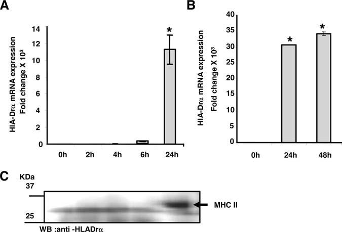
IFN-γ stimulated SMCs express great levels of HLA-Drα. RNA was extracted from human aortic SMCs that were treated with or without 100 U/mL IFN-γ at various times (2, 4, 6, 24, and 48 hours). TaqMan probe and specific primers were used to amplify cDNA for HLA-Dra (A and B) by real-time PCR. Each experiment was repeated at least 3 times and values represented as mean±SD. The data were evaluated for significance by ANOVA followed by Sheffe's post hoc analysis using SPSS software (*P<0.01). C, Cytoplasmic proteins were extracted from human aortic SMCs that were induced with IFN-γ (100 U/mL) at different times (2, 4, 6, and 24 hours). Proteins (20 mg) were separated in 10% SDS gel and electroblotted to a nitrocellulose membrane. The membrane was incubated with HLA-Dra antibody (FL-254; Santa Cruz Biotechnology) that detected a correct size band of 32 kDa after 24 hours of IFN-γ treatment. The chemiluminescence was measured with a Kodak Image station.