
Fig. 6.
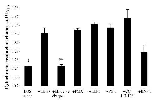
Cytochrome c reduction by ROS was enhanced by AMP and endotoxin. THP-1 cells primed with 5.6 pmole ml−1 of N. meningitidis LOS in presence of 2 μg ml−1 of cationic peptides LL-37, LLP1, PMX, CG117-136, PG-1 and HNP-1 overnight. Respiratory burst was triggered with 100 ng ml−1 PMA in presence of 90 μg ml−1 of freshly dissolved cytochrome c with or without 250 units of SOD. LL-37-ve is negatively charged peptide used as control. Cytochrome c reduction by the generated ROS was recorded as the absorbance of reduced cytochrome c and the change in OD at 550 nm. Each bar represents the mean of triplicate readings after 10 min of incubation. This experiment is representative of four different experiments. P-values calculated for AMP + LOS compared with (Student t-test) to LOS alone* or to LOS + LL-37-ve charge** and were < 0.001.