
Fig. 5.
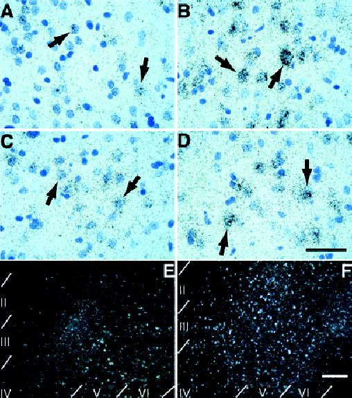
Emulsion autoradiography of forebrain cortical regions hybridized to arc antisense probe. (A and B) Silver grains in layer V/VI of the prelimbic regions of the prefrontal cortex accumulate preferentially over cells with lightly stained, large nuclei (arrows in A and B) in a rat exposed to nicotine cues (B) compared with saline cues (A). This silver grain pattern is consistent with neuronal localization of induced arc (scale bar, 50 μm for A–D). (C and D) Silver grains in layers V/VI of the lateral orbital prefrontal cortex also accumulate preferentially over cells with lightly stained, large nuclei (arrows in C and D) in a rat exposed to nicotine cues (D) compared with saline cues (C). This silver grain pattern is again consistent with neuronal localization of induced arc. (E and F) Lower power magnification, dark-field photomicrographs of sensorimotor cortex (corresponding to Fig. 4I and J). Recruitment of cells expressing arc occurs preferentially in layers II–IV in the sensorimotor cortex of rats exposed to nicotine cues (F) compared with rats exposed to saline cues (E). This laminar pattern of expression was evident in most cortical areas analysed (scale bar, 200 μm for E and F).