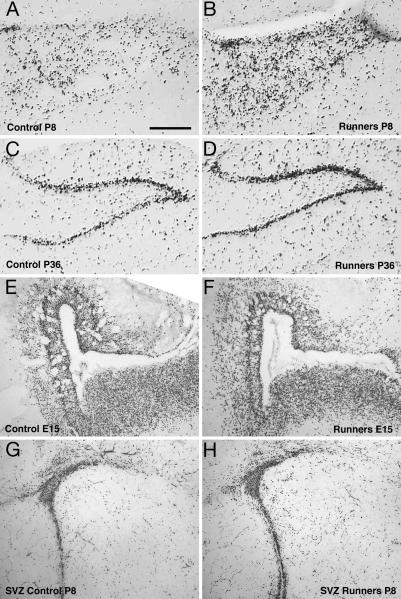
Fig. 2.
Proliferating cells in the dentate gyrus of RUN and CTR pups at P8 and P36. The representative micrographs show an anti-BrdU staining in the granule-cell layer of the dentate gyrus. (Scale bar, 250 μm.) (A and B) At P8, proliferating BrdU-labeled cells were dispersed over the granule-cell layer in RUN (n = 6) and CTR (n = 8). RUN had 71% more BrdU-labeled cells than CTR (P = 0.01). (C and D) At P36, most of the BrdU-labeled cells were detected in the characteristic cell band of the granule-cell layer. Also, at this time point, the total number of granule cells of RUN (n = 5) was 140% of CTR (n = 6), P = 0.001. (E and F) At E15, in RUN, a reduced number of proliferating (i.e., BrdU-labeled) cells in the hippocampal anlage and the adjacent ventricular and SVZ was found. This difference is not obvious in the photomicrograph but was statistically significant upon quantification (see Fig. 1C). (G and H) At P8, no obvious difference (as in the hippocampus, see A and B) in the number of BrdU-labeled cells was found in the SVZ. However, the experiment was not designed to study the SVZ quantitatively, and no strong conclusions should be drawn from the impression gained from these images.

