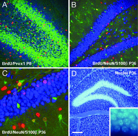
Fig. 3.
Confocal microscopic and DNA stain bisbenzemide analysis of granule cells and astrocytes in the dentate gyrus (DG) of pups from running mothers (RUN). Within each panel, the antigens investigated are listed in the order red, blue, and green. (A) Distribution of granule cells in the dentate gyrus at P8 was identified by their expression of the neuronal transcription factor Prox1. Newly generated granule cells were identified by immunoreactivity for proliferation marker BrdU (red) and Prox1 (blue). One day after BrdU application, the number of Prox1-expressing newly generated granule cells increased significantly in RUN compared with CTR. (B and C) At P36, survival of newly generated cells was quantified by their incorporation of BrdU (red). Neurons and astrocytes were characterized by their expression of NeuN (blue) or S100β (green). The survival rate of new neurons and astrocytes was significantly increased in RUN compared with controls. (D) Total granule-cell numbers were determined by using stereology (optical fractionator) based on DNA stain bisbenzemide (Hoechst 33258; see inset for higher magnification). Increased neurogenesis in the DG in RUN resulted in an absolute increase in the total number of granule cells at P36. [Scale bar in D for all panels; 100 μm (A and B), 30 μm (C), and 200 μm (D).]

