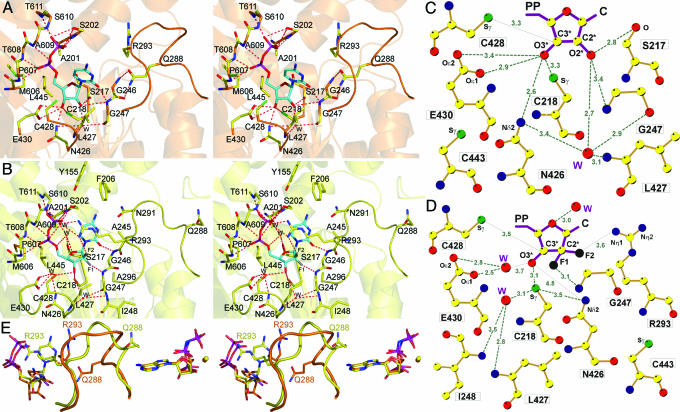
Fig. 2.
Catalytic-site interactions. (A) Stereoview of CDP (orange). Interacting atoms: oxygen, red; nitrogen, blue; phosphate, magenta; sulfur, green; substrate carbons, cyan; protein non-Cα carbons, yellow; Cα carbons, as secondary structure, orange. (B) Stereoview of GemdP. Interacting atoms are colored as in A, except that sulfur is orange; Cα carbons, as secondary structure, are yellow; and fluorines are gray. (C) Ligand plot of CDP ribose interactions. Colors are as in A, except that carbons are yellow. (D) Ligand plot of GemdP interactions. The van der Waals contact to L427 is omitted for clarity. (E) Stereoview of loop-2 superposition of AMPPNP–CDP (orange) and AMPPNP–GemdP (yellow). Substrate/inhibitor is seen on the left, and the effector is on the right. The color scheme is the same as in C, but fluorine is black.

