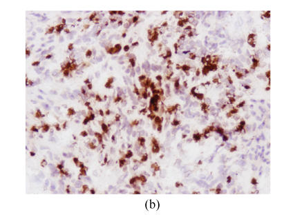
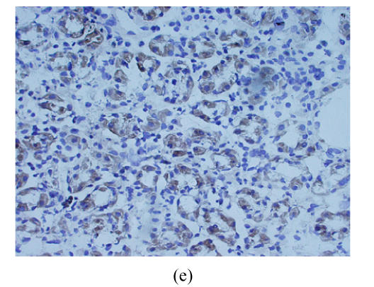
Fig. 2.
Cryostat section of gastric carcinoma tissue (magnification 200×). Positive staining of CD97 and CD55 was observed on cytoplasm and membrane of gastric carcinoma cells and showed brown-yellow, the cellular nuclears generally showed blue. When the staining was too strong, the cellular nuclears was covered by brown-yellow (a) Strong CD97stalk-staining was localized in scattered tumor cells surrounded in matrix or small tumor cell clusters; (b) Strong CD97stalk-staining was observed on cytoplasm of signet ring cell carcinomas; (c) Strong CD55-staining was located on cytoplasm of signet ring cell carcinomas; (d) CD55-staining was located on glandular luminal sides, and was expressed over the entire surface where the ducts were present; (e) Positive-staining of CD55 was frequently observed in normal gastric glandular epithelium


