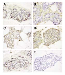
Figure 2.

Immunohistochemical analysis of TGF-β expression in the rat mammary gland. The first thoracic (number 2) mammary gland was harvested from 15-week-old rats that had been treated with NMU at age 8 weeks. Sections were immunostained (see Materials and method), with the following antisera: (a) anti-TGF-β1-LC (stains predominantly intracellular TGF-β1); (b) anti-TGF-β1-CC (stains extracellular TGF-β1); (c) anti-TGF-β2; (d) anti-TGF-β3; (e) anti-LTBP; and (f) normal rabbit immunoglobulin. The brown stain indicates a positive immunoperoxidase reaction. Images were shot at 630× original magnification.
