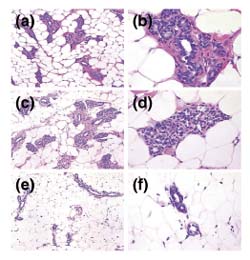
Figure 4.

Treatment with tamoxifen affects the histology of the rat mammary gland. Representative hematoxylin and eosin stained sections of the first thoracic gland of 15-week-old rats that had undergone the following treatments: (a, b) No treatment; moderate numbers of mammary gland lobules are present containing primary, secondary and tertiary ductules, as well as developing alveoli. (c, d) Initiation with NMU at 8 weeks of age; no significant histologic differences are noted in mammary gland development from that in untreated control animals. (e, f) Initiation with NMU at 8 weeks, followed by treatment with tamoxifen from 9 to 15 weeks of age; scant numbers of atrophic primary and secondary mammary gland ductules are noted, with no alveolar bud development evident. (a, c, e) Shot at 100×; and (b, d, f) shot at 400× original magnification.
