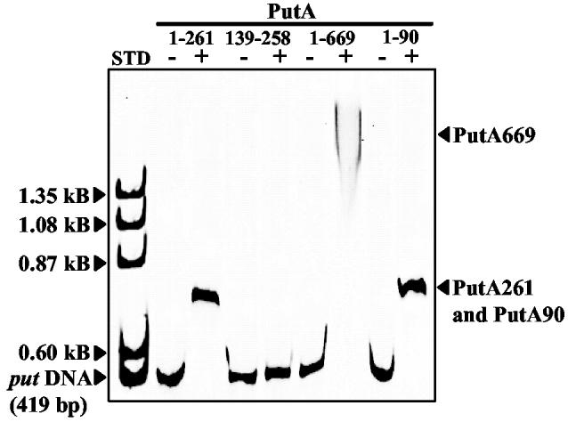
Fig. 1.

Gel mobility shift assays of truncated PutA proteins and the put control DNA. PutA261, PutA139-258, PutA669 R230A/R234A, and PutA90 (1 μM monomer each) were incubated in separate binding mixtures (50 mM Tris, pH 7.5) with IRdye-700 labeled put control DNA (5 nM) at 20 °C. The complexes were separated using an agarose/polyacrylamide (0.5%/3%) native gel at 4 °C. The position of uncomplexed put control DNA and the protein-DNA complexes are indicated. The left-hand lane (STD) shows the migration of uncomplexed put control DNA (0.419 kB) and ΦX174 ladder DNA (500 ng) with molecular size standards of 1.353 kB, 1.078 kB, 0.872 kB and 0.603 kB. The gel was stained with ethidium bromide to visualize the molecular size standards.
